The eye is an organ of vital importance and quite complex since it is directly related to structures that connect with the brain. The eye captures light and converts it into nerve impulses that travel through the optic nerve to the brain for interpretation. But to understand how the eye works, we must first know what its function is.

HOW DOES THE VISUAL SYSTEM WORK?
Different parts of the eye work together to help us see.
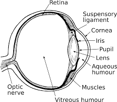
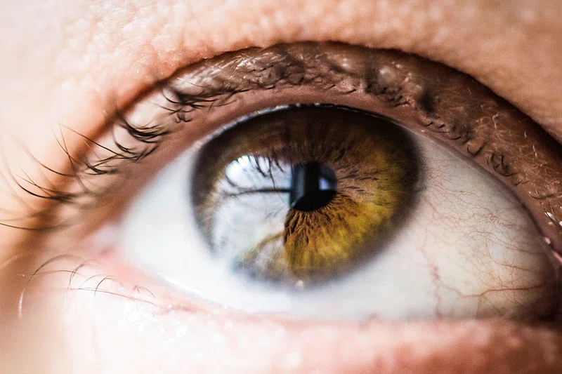
The part of the light that enters the eye through the opening is called the pupil. The iris (the colored part of the eye) controls the amount of light the pupil receives.
First, light passes through the cornea (the transparent surface of the eye). The cornea is shaped like a dome and deflects light to the crystalline, and from the crystalline light is focused on the retina.
When light reaches the retina (the layer of light-sensitive cells at the back of the eye), special cells called photoreceptors convert the light into electrical signals.
The electrical signals travel from the retina to the brain. The brain then converts the signals into images that we see.
The visual system is very complex and each structure that forms the eyeball and the visual system in general has a function and a vital importance. The image that we can see is thanks to the small functions that each part of the eyeball does. For example, such a simple part as tears, is of vital importance to lubricate the eye. If the eye is not moisturized, this will affect the cornea and all the structures of the visual system so there will be no vision.
EXTERNAL PARTS OF THE EYE
- Eyelids: this is the structure in charge of protecting the eyeball. There is the lower and upper eyelid, and both are made up of two parts, the external and the internal.
- Eyelashes: they also have a protective function and are found in both the upper and lower eyelids.
- Eyeball: it is what we usually say “eye”. It is more or less spherical and is located inside the orbital cavity. It weighs about 7g and measures approximately 2.5 cm. It is composed by the outer layer: forms the upper part of the eyeball. It is formed by structures such as the cornea, the anterior part of the sclera and the conjunctiva. The middle layer: formed by the uvea, the iris and the sclera. The inner layer: it is formed by the retina.

EYEBALL PARTS
⦁ Sclera: the white, fibrous layer of the eye. It is composed mainly of collagen fibers. It extends from the cornea (anterior part) to the optic nerve (posterior part).
⦁ conjunctiva: is the transparent membrane that lines the sclera and the inner side of the eyelids.
⦁ cornea: transparent layer that covers only the iris, pupil and anterior chamber of the eye. It is in contact with the tear film.
⦁ tear film: set of three layers (outer, middle and inner) responsible for covering and protecting the eyeball.
⦁ pupil: black circular dot in the center of the iris. It is a hole that regulates the entry of light into the retina.
⦁ anterior chamber: space filled with a liquid called aqueous humor that lies between the iris and pupil and the cornea.
⦁ posterior chamber: communicates the iris with the crystalline lens.
⦁ crystalline lens: lens behind the iris and aqueous humor and in front of the vitreous humor. It has a high protein content. It is responsible for refracting light coming from outside to form the image on the retina. It deflects the light rays in such a way that it produces a visible image on the retina, and from there, the image is sent directly to the brain in the form of sensors through the optic nerve. The lens is able to bulge to achieve good near and far vision, which is achieved by increasing or decreasing the curvature and thickness, in a process called accommodation, the lens adjusts vision with the help of the ciliary muscle.

⦁ iris: it is between the cornea and the crystalline lens.made up of muscle fibers that dilate or contract depending on the light that penetrates the eye.
⦁ retina: membrane located at the back of the eye. Receives light from the outside to send it through the optic nerve to the brain. It consists of 3 vital parts for vision the macula: it produces the central vision. It allows us to see colors and details. the fovea: it is the center of the macula where the rays coming from the outside focus, it is the center of vision. Optic papilla: it is the beginning of the optic nerve where all the neurons are grouped to transmit the nerve impulse through the optic nerve.
⦁ optic nerve: transmits information from the retina to the brain. It is a nerve that has fibers in the papilla part of the retina. It receives the images that it sees from the outside to send information to the brain through a chemical and electrical process that converts electrical impulses into images. It plays a very important role in vision, so if the optic nerve is damaged, there is no vision.
⦁ aqueous humor: transparent liquid that forms in the ciliary body. It circulates and nourishes the avascular structures of the eye.
⦁ vitreous humor: a clear liquid formed mainly of water located between the retina and the lens.
⦁ ciliary body: extension located between the choroid and the iris is responsible for producing aqueous humor. It is composed of the ciliary muscle, ciliary epithelium and ciliary processes.
⦁ choroid: membrane located between the retina and the sclera and is responsible for supplying nutrients to the internal structures of the eye.
Related Posts

defective curvature of the cornea or lens of the eye

ANATOMY OF THE EYE


